- Home
- Types of Radiation
The external beam radiation therapy is the form of radiation treatment, delivering several beams of high-energy photons, electrons or protons through the skin to the cancer site and the immediate surrounding area in order to destroy the cancer cells. Typically the patient lies on a couch and the Gantry or Head of the machine rotates around, delivering radiation at a particular part of the body. While megavoltage photons or protons are used to treat more deep-seated cancers (e.g. bladder, bowl, prostate, lung or brain),Electron beams are used for treating skin cancer and cancers seated in superficial tissues.
Steps in External Radiation Treatment

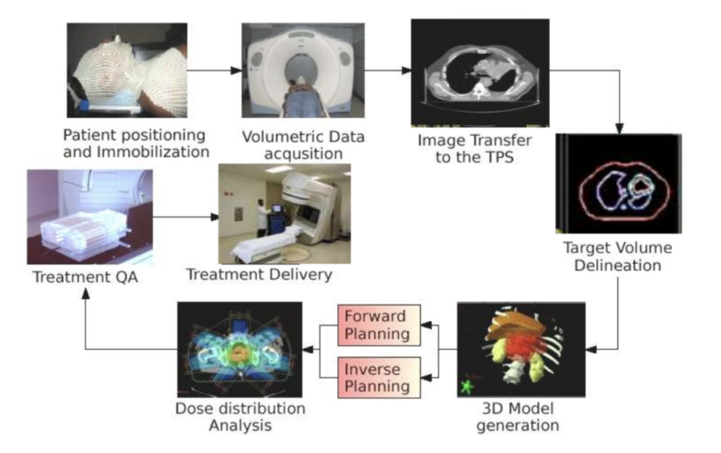
Simulation
Simulation is the most important preparation step before the actual radiation treatment.
Siemens Somatom Sensation-Open CT- Simulator The simulation room is equipped with this dedicated large-bore CT scanner called as Simulator.
During simulation,–
1.The treatment setup is replicated by positioning the patient on the flat couch immobilized by specially designed devices.
- The patient is then aligned with the help of reference low-energy lasers in the room and marked on the skin with tattoos.
3.Finally, a CT scan is performed to acquire the anatomy of the reference treatment site.
- This CT scan would be used to identify the tumour area and also the surrounding normal critical organs to create a treatment plan that will guide the treatment machine to target the tumour accurately and spare the normal organs.
- The simulated setup will be exactly reproduced before each treatment by matching the reference lasers in the treatment room to the tattoos and comparing the 2D/3D on-board images with the simulation CT scan. The patient may need to spend from 15 minutes to 45 minutes to complete the entire process of simulation.
A change of planning during the course of the treatment may often be need after a few days of radiation for selected cases using this equipment .
Linear accelerator, or LINAC,—
This is a machine that is commonly used to deliver external beam radiation treatments ( EBRT) to cancer patients. A linear accelerator is programmed to deliver high-energy X-rays that conform to the specific size, shape and location of a tumor. In this way, the LINAC can target and destroy cancerous cells in a precise area of a patient’s body with minimal exposure to the surrounding healthy tissue.
The Radiation Oncology Department is equipped with 4 LINACs including , Accuray- TomotherapyRadixact X9 with 6FFF photon beam with MV Imaging, capable of advanced state of arts IMRT, IGRT, Volumetric therapy and TBI techniques, Varian – Clinac-iXRapid Arc, Varian – True Beam Stx,Elekta -Synergywith facilities of Photon Beam, Electron Beam, providing
- 3D-Conformal Radiotherapy,
- Intensity Modulated Radiotherapy (IMRT),
- Image Guided Radiotherapy(IGRT),
- Volumetric Modulated Arc Therapy (VMAT)
- Tomotherapy
- Stereotactic Radiotherapy and (SRT)
- Stereotactic Radiosurgery(SRS).
- 3D-Conformal radiotherapy–3D CRT, or three-dimensional conformal radiation therapy, is an advanced technique for radiotherapy, that incorporates the use of imaging to generate three-dimensional images of a patient’s tumor and the nearby organs. This technology distinguishes 3D CRT from older forms of conventional radiation therapy
- Intensity Modulated Radiation Therapy – IMRT
Intensity Modulated Radiation Therapy (IMRT) is an approach that delivers a high, conformal dose to the tumor while restricting the exposure to surrounding normal tissue.
The benefits of IMRT
The benefits of IMRT include:
- IMRT delivers higher doses of radiation for destroying the tumor, regardless of size, shape or location, thus enabling a higher chance of cure.
- IMRT uses computer-generated images to plan and delivers tightly focused radiation beams, thereby sparing surrounding healthy tissue. Hence side effects are minimized.
- IMRT may make re-treatment with radiation possible, since healthy tissue initially received less radiation than with conventional therapy. The radiation beams are planned to avoid critical structures.
- IMRT brings the advantages of radiation therapy to a wider range of cancer patients, including patients with difficult-to-reach tumors or tumors located close to vital organs.
IMRT uses multiple small radiation beam lets of varying intensities to precisely conform to a tumor. The radiation intensity of each beam is controlled, and the beam shape changes throughout each treatment using multi leaf collimators, allowing a relatively uniform dose to the tumor and a sharp fall-off of the dose to surrounding normal organs.
- Image Guided Radiation Therapy – IGRT
The Department is equipped with “Image Guided Radiation Therapy (IGRT)”
IGRT combines the traditional radiation treatment with a simultaneous cone beam CT scan to allow for greater precision and accuracy when targeting and treating tumors.
Frequent two and three-dimensional images are taken during a course of radiation treatment, to ensure that the treatment volume is being accurately targeted.
The images are taken after the patient is set up for treatment. Once the image is acquired it automatically fuses with the planning CT. Upon fusion of the two images sophisticated software is able to visualize whether or not the treatment volume is off target from the planned target and make the necessary adjustments to ensure accurate treatment.
In conventional radiation treatment, the area at risk is targeted and a margin is added around it to allow for changes in the patient’s position and potential movement of the target area. Using IGRT, margins can be minimized, thus sparing the normal tissue that does not otherwise need to be treated.
IGRT has opened the door to true four dimensional (4D) radiation treatment. In addition to dealing with the three dimensions of space, IGRT deals more effectively with the issue of tumor motion in time – the fourth dimension using 4D imaging, 4D simulation, 4D treatment planning, 4D treatment delivery and 4D verification.
The active breathing control is such a device. Because inhaling and exhaling can move organs as we breathe, it may be difficult to aim radiation at a tumor site without involving nearby organs. This is particularly so in the abdomen and chest areas. An active breathing control device spares nearby organs unnecessary radiation and providesclinicians with an accurate method for delivering radiation.
4.Volumetric Modulated Arc Therapy
Volumetric Modulated Arc therapy (VMAT) is a novel addition to our radiation department for treatment of cancers. The use of fast sweeping uninterrupted arc of radiation beam not only reduces the time of the treatment from 8 – 15 minutes to less than 3 – 5 minutes but also enables in dose reduction to the patient without compromising the efficacy of the treatment. It enables the clinicians in creating 3D sculpted dose volume structures with optimal beam delivery providing high target conformance with safe-guarding surrounding normal tissues. With faster delivery and lesser treatment times, it enables the clinicians to counter act the element of organ motions; with faster treatment time chances of geographical miss decreases rapidly.
- Tomotherapy —Tomotherapy is an advanced treatment for cancer that combines the precision of intensity-modulated radiation therapy (IMRT) with the real-time accuracy of CT scanning or or IGRT. The benefits of Tomotherapy include:
- Improved monitoring of tumor position and size in real-time.
- More targeted radiation shaped to thetumor.
- Higher doses of radiation to the tumor, potentially increasing the efficacy of treatment.
- A markedly reduced amount of radiation reaches healthy organs and tissue next to the tumor. This helps reduce side effects and long-term complications.
This is ideal for tumors that are hard to reach, or next to vital organs.In addition, it can treat multiple tumors at one time.
- Newer Technologies In Radiation Oncology
- DIBH—
Deep inspiration breath hold (DIBH) is a technique that takes advantage of a more favorable position of the heart during inspiration to minimize heart doses over a course of radiation therapy.When one takes a deep breath and holds it, her diaphragm pulls the heart away from the chest. This is known as a deep inspiration breath hold (DIBH).The radiation is delivered to the breast while the patient is holding her breath deeply for 20 seconds. This provides protection for the heart.This benefit is the greatest in those patients with left-sided disease and those receiving IMC irradiation. DIBH has also been shown to decrease dose to the lungs.
- Stereotactic Radiosurgery
Stereotactic radiosurgery is the use of multiple small beams to deliver very high ablative doses of radiation, from different angles and planes, shaped to the size of the tumour. It is most often used on small, well-defined tumours. Three-dimensional imaging is used to determine the exact coordinates of the tumor to localize it. Contrary to its name, it is a non-surgical radiation therapy that ca be used as an alternative to invasive surgery.
Its biggest benefits over conventional therapy are:
- It can treat very small tumors or those located in hard-to-reach places.
- Treatment times are much shorter.
Different kinds of stereotactic radiosurgery may be used, depending on the type of cancer and where it is located in the body. Stereotactic radiosurgery is used to treat brain and spinal cancers.
SBRT PLAN
Stereotactic body radiation therapy (SBRT) offers the same benefits as stereotactic radiosurgery and is used to treat similar tumours at extracranial sites like the head and the neck, Liver, Pancreas, Bones and any other sites needed.
Brachytherapy
Brachytherapy means delivering radiation from a short distance as by keeping radiation close to tumour.
High dose rate brachytherapy
Brachytherapy by this technique involves short treatment time as dose in delivered at a high dose rate (HDR). This is a special device which usually allows outpatient treatment and there is no need for hospitalization of the patient. There is no risk of exposure of radiation to oncologists, physicist, technicians and visitors close to the patient. Generally, it is used for treatment of cancers of the cervix, Oral Cavity, Breast, Lung, Prostate, skin and soft tissue sarcomas.
HDR Brachytherapy uses dose rates results in treatment times of few minutes. The treatment be used alone or in combination with external beam radiation.
Brachytherapy Room
Brachytherapy Applications
- Intracavitary Brachytherapy
It consists of positioning applicators (with or without the radioactive sources) into a body cavity in close proximity to the target tissues . The most widely used intracavitary treatment technique is insertion of tandem and colpostat applicators for treatment of cervix cancer.
- Interstitial brachytherapy
It consists of surgically implanting small radioactive sources directly into the target tissues through either stainless steel needles all polyvinyl catheters, which are inserted in to the lumen. The procedure is commonly used in cancer breast, soft tissue sarcoma, cancer prostate, cancer cervix, head & neck cancers and at any other sites which can be approached for implantation. A permanent interstitial implant remains in place forever. Iridium wire implants are commonly used in the management of carcinoma breast and peripheral soft tissue sarcomas. Permanent gold, iodine or palladium seed implants are used for cancer of the prostate, lung and anal canal.
- Intraluminal Brachytherapy
It consists of inserting a single line source into body lumen so as to treat its surface and adjacent tissues. Commonly treated sites are cancers of the lung and oesophagus.
- Mould Therapy
It consists of an applicator containing an array of radioactive sources that is usually designed to deliver a uniform dose distribution to skin or mucosal surface. It is commonly used for skin cancers over curved surfaces like hands, lips, legs, ears and recurrences in carcinoma breast.


